

Gastric ulcer biopsy Download Scientific Diagram

gastric ulcer biopsy Knowledge Shenzhen Manners Technology Co Ltd

Biopsy of the caecal ulcer The colic biopsy shows an acute

A B Biopsy of the ulcer revealed chronic active inflammation of the

A B Biopsy of the ulcer revealed chronic active inflammation of the

Ulcer biopsy results by year Download Scientific Diagram
57+ Images of What Is Ulcer Biopsy

Gallery of What Is Ulcer Biopsy :

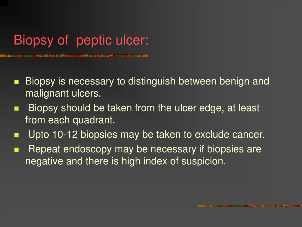
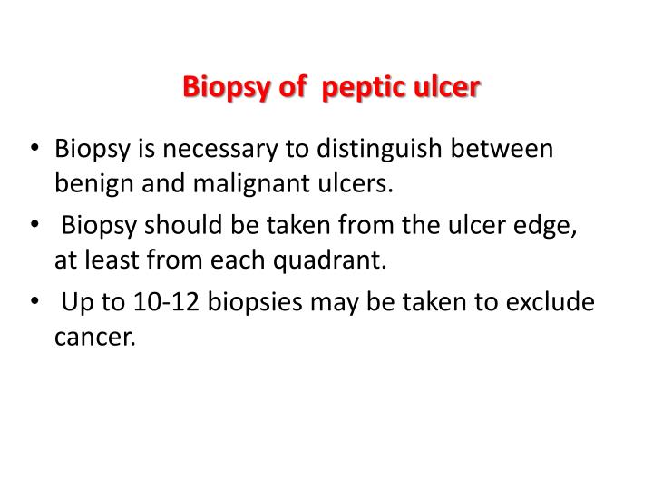
When to Biopsy an Ulcer

Photomicrograph of biopsy notable for ulcer secondary to

Biopsy of stomach Biopsy of the antral ulcer in the stomach shows

The pathological findings of the biopsy of the ulcer at the

Clinical features prior to initial biopsy A 1 2 cm x 0 6 cm ulcer with

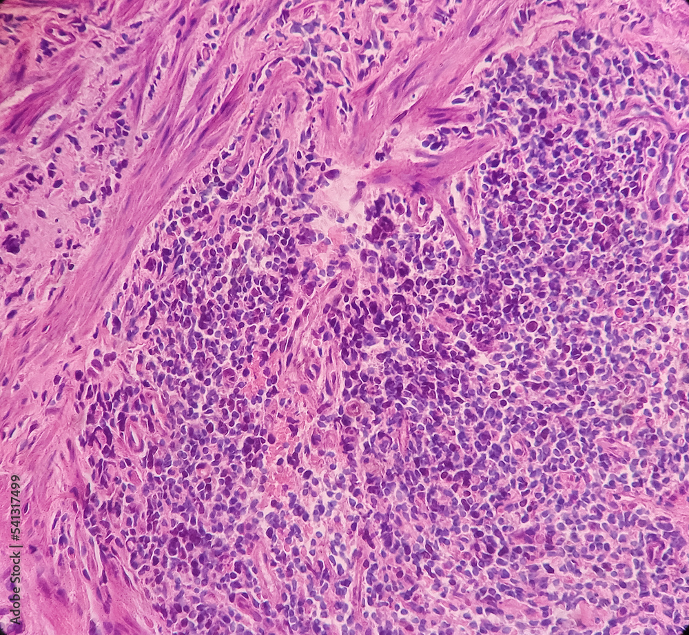
Histopathology slides of the ulcer biopsy with hematoxylin and eosin

Histopathology slides of the ulcer biopsy with hematoxylin and eosin

Stomach ulcer biopsy showed a dense infiltrate composed of large

a and b Biopsy from the ulcer edge in the skin and transverse colon

Representative images of biopsy samples from a duodenal ulcer

Representative images of biopsy samples from a duodenal ulcer

Representative images of biopsy samples from a duodenal ulcer

Gastric biopsy of non bleeding ulcer Poorly differentiated

Biopsy of ulcer at A a lower magnification showing squamous mucosa

Biopsy from the ulcer site with chronic active gastritis and intestinal
Biopsy of the border of the ulcer A Tumour invasion is observed

Histological analysis of the gastric ulcer biopsy showing malignant

Leg Ulcer Pictures 2024 CACVI

Gastric ulcer biopsy at 4 215 magnification A Hematoxylin and Eosin

Biopsy of ulcer at A a lower magnification showing squamous mucosa
Gastric biopsy of non bleeding ulcer Poorly differentiated
Biopsy of ulcer at A a lower magnification showing squamous mucosa
Biopsy from the ulcer site with chronic active gastritis and intestinal
Biopsy of the border of the ulcer A Tumour invasion is observed
Histological analysis of the gastric ulcer biopsy showing malignant
Leg Ulcer Pictures 2024 CACVI
Gastric ulcer biopsy at 4 215 magnification A Hematoxylin and Eosin
Biopsy of ulcer at A a lower magnification showing squamous mucosa

Microscopic view of the biopsy specimen obtained from ileal ulcer

High and low power view of duodenal ulcer biopsy show collections of

Photomicrograph of the oral biopsy A non specific ulcer with

Photomicrograph of the oral biopsy A non specific ulcer with

Microscopic features of the re biopsy specimen a Ulcer and severe

Biopsy PPT

Histopathology of the biopsy from the leg ulcer showing nonspecific

Anatomopathological examination of skin ulcer biopsy showing infi

Photomicrographs of the ulcer skin biopsy A Acanthotic epidermis

Gastric ulcer biopsy Section shows fragments of gastric mucosa The
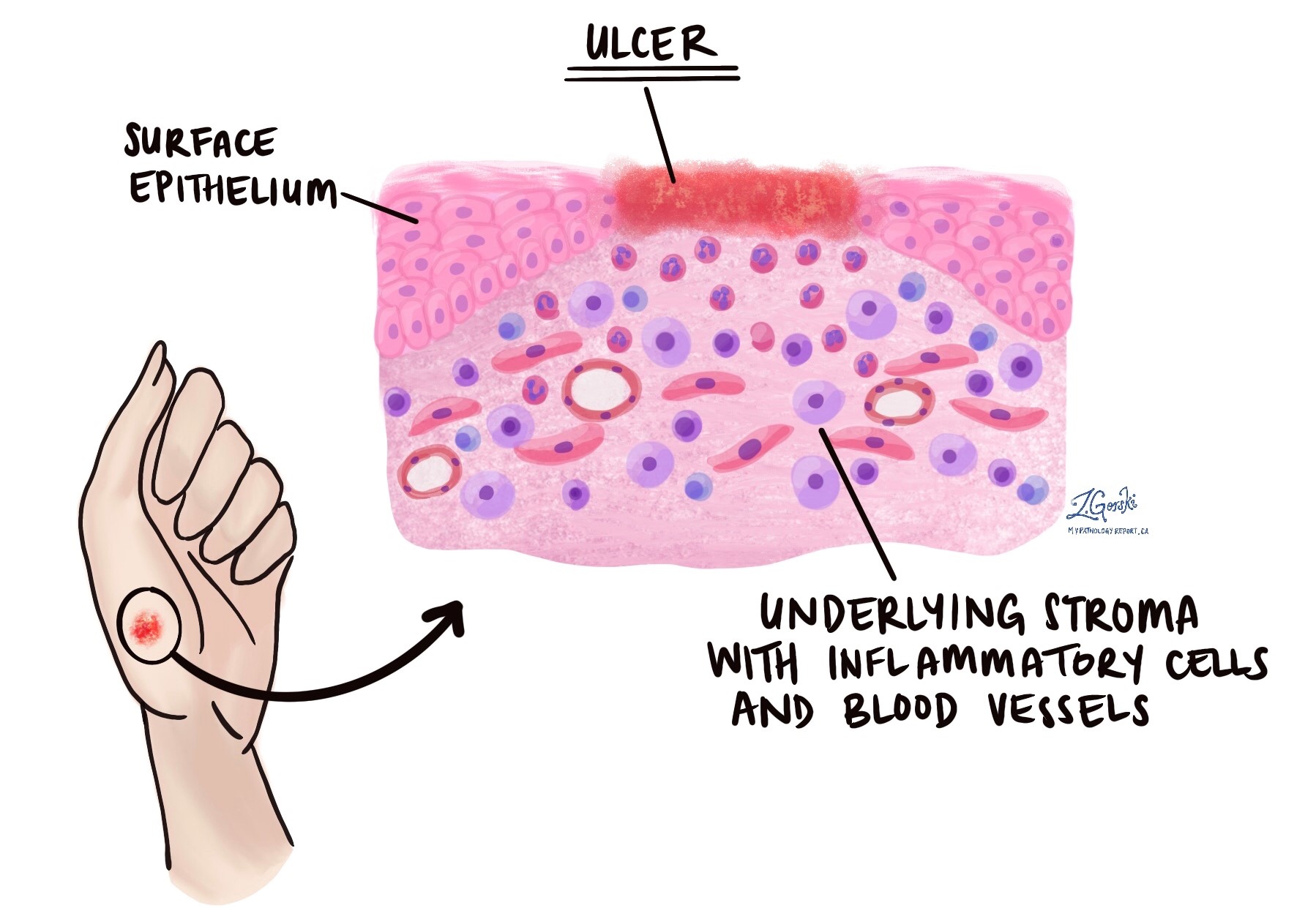
Ulcer MyPathologyReport ca

Histological analysis of the gastric ulcer biopsy showing malignant

A scanner view of a non specific ulcer edge biopsy showing a

Histologic examination of biopsy specimen from the ileal ulcer

Microscopic features of the re biopsy specimen a Ulcer and severe

Peptic ulcer

Biopsy of ulcer performed on day 4 H amp E stain 400 194 a and PAS stain

Anatomopathological studies of skin biopsy On the left there is a

Biopsy from the edge of one of the ulcers H amp E 100 215 magnification

Endoscopy The ulcer is a plain superficial ulcer located by the major
Biopsy from the edge of one of the ulcers H amp E 100 215 magnification
Endoscopy The ulcer is a plain superficial ulcer located by the major

Endoscopy The ulcer is a plain superficial ulcer located by the major

PEPTIC ULCER ppt

Photomicrograph of biopsy specimens A and B Biopsy specimens of

PPT PEPTIC ULCER disease PUD PowerPoint Presentation free download

Endoscopic view of the ulcer in the fundus of the stomach The arrow

Malignant looking ulceration one week post incisional biopsy

Finding after mass biopsy of the ulcerative lesion Download
PPT PATHOLOGY AND PATHOGENESIS OF PEPTIC ULCER PowerPoint

Differences between ulcers in patients subjected or not subjected to a

Skin biopsy findings showing ulceration likely secondary to

Upper gastrointestinal endoscopy revealing a 1 5 194 1 5 cm and big ulcer

One week postoperative photograph after excisional biopsy Download

Left Initial macroscopic appearance of the ulcers Middle Biopsy of
PPT PEPTIC ULCER disease PUD Pathophysiology PowerPoint

Microscopic examination of the biopsy A reveals the edge of an
Anal canal ulcer biopsy Chronic nonspecific proctitis show anal

Endoscopic biopsy specimens showing ulcers lined by epithelioid

a Skin ulcers on left leg b skin biopsy H and E 215 100 low power

Figure 2 from Mapping Biopsy Procedure on Management of Severe Buruli

Subsequent biopsy of the ulcerated lesion in case 1 It shows a

Pathology images Biopsy from the gastric ulcers showed the

Pathology images Biopsy from the gastric ulcers showed the

Biopsy histopathology of the lesion in case 4 showing partly ulcerated
Gastric Ulcer Histology

Keith Siau on Twitter quot Throwback to this landmark study In summary

Peptic ulcer Stock Image M280 0185 Science Photo Library
What Is Ulcer Biopsy - The pictures related to be able to What Is Ulcer Biopsy in the following paragraphs, hopefully they will can be useful and will increase your knowledge. Appreciate you for making the effort to be able to visit our website and even read our articles. Cya ~.
