

Number of severe and mild MRI complications observed for 66 MRI scans

Pin by phuong hoai on MRI Medical radiography Radiology imaging
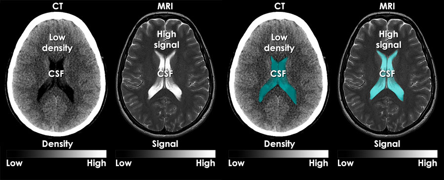
MRI interpretation What are MRI images

What Does an MRI Show and How Does It Work Ezra

MRI Findings NBIA

MRI interpretation Systematic approach
61+ Images of What Does Mild Mean On Mri

Gallery of What Does Mild Mean On Mri :

Patient s first MRI which revealed a mild enlargement and asymmetric

On magnetic resonance imaging MRI a lesion appeared mild

What do my MRI results mean

Understanding MRI Medical Articles Hub

4 Common MRI Misconceptions Explained

21 M just had an MRI done what does this mean r backpain

Your MRI Matters Advantage Health Group
Why does it take so long for an MRI scan AIRS Medical Inc

Principles of MRI PPT

MRI imaging manifestations of some patients and healthy people a An
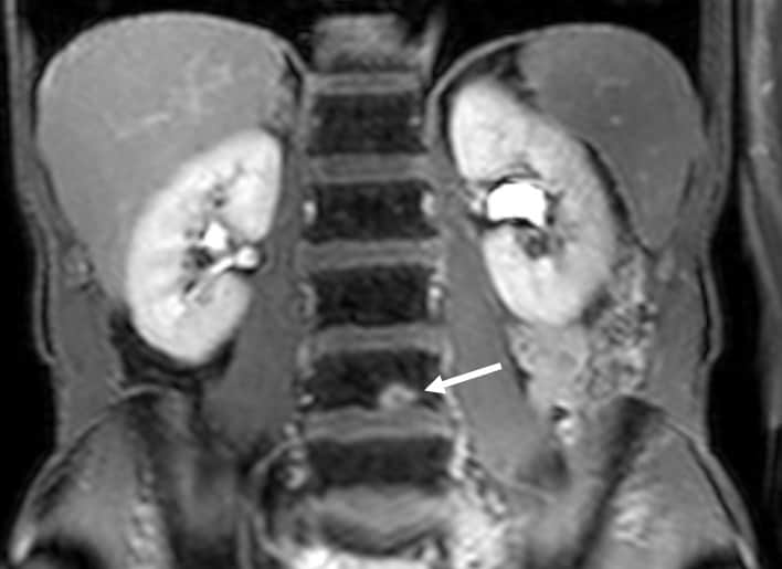
DIAGRAM Diagram Of Mri MYDIAGRAM ONLINE

Hello How bad the MRI looks The symptoms are mild r HerniatedDisc

Hello How bad the MRI looks The symptoms are mild r HerniatedDisc

Hello How bad the MRI looks The symptoms are mild r HerniatedDisc

Left a b Conventional MRI showing normal findings while c d

The description of MRI findings Download Scientific Diagram

MRI images of the patient A F Preoperative MRI examination revealed

Understanding Your MRI MS Living Well

MRI image of the patient A B Day 7 of symptom onset Both A and
MRI T2 weighted MRI shows a low signal lesion identified as an air
Understanding Your MRI MS Living Well
MRI image of the patient A B Day 7 of symptom onset Both A and
MRI T2 weighted MRI shows a low signal lesion identified as an air

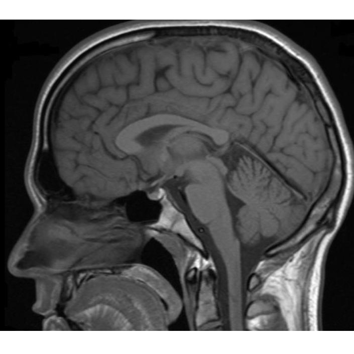
T 1 weighted MRI In the acute period the MRI scan is normal A

T 1 weighted MRI In the acute period the MRI scan is normal A

The MRI at 50 what lies ahead

Mri question difference between images New diagnosis and before

Mri question difference between images New diagnosis and before

1 MRI interpretation docx

What Do Your MRI Results Really Mean InfoPhysiotherapy
MRI CancerQuest

Typical MRI findings in a PA patient F 26 years a T1 weighted MRI

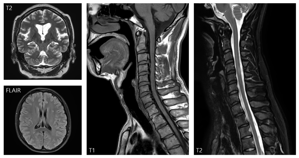
The Basics of MRI Interpretation Radiology Geeky Medics
Body MRI reinterpretations plagued by discrepancies and errors

MRI images and clinical symptoms before and after the procedure

MRI images and clinical symptoms before and after the procedure

The MRI at 50 what lies ahead

Different types of MRI

MRI appearance of normal and pathologic features

MRIs of the patient A Enhanced T1 weighted MRI taken 23 days after

MRI results in our patient in 2019 a b and 2021 c d a

a Axial T1 weighted MRI image showing low signal intensity poorly

Patient s MRI performed at the age of 8 months Showing no

Axial T1 weighted MRI image A shows a well defined heterogeneous low

T2 weighted MRI scan showing normal appearances 1 year after symptom

Coronal view of a T1 weighted MRI showing an abnormal elongated round

Do these MRI pics look normal r DiagnoseMe

MRI of male patient aged 22 years old showing normal radiological
T2 weighted MRI scan showing normal appearances 1 year after symptom
Coronal view of a T1 weighted MRI showing an abnormal elongated round
Do these MRI pics look normal r DiagnoseMe
MRI of male patient aged 22 years old showing normal radiological

The ML appearance in MRI is normal Download Scientific Diagram

FIGURE E A The example of MRI positive images of a a year old female

MRI with and without contrast in seven sections indicating A interval

Figure MRI images at presentation and at 6 month follow up A
MRI scan Tl weighted image sagittal section near midline revealing

Spectrum of MRI abnormalities in pre symptomatic patients A Faint
MRI in patient 1 at the time of diagnosis upper arrow and 4 months

What is this on my MRI AskDocs

Images from MRI performed at 15 month A C and at 46 month of age
The preoperative axial MRI shows an area of a low intensity signal on a

Is this something significant Report says normal but I found this
Incidental Finding on MRI in a 22 year old man Manual of Medicine

Image features of the MRI variables a d Four grades of relationship

MRI examination of the patient 1 5 h after the onset of symptoms A

MRI images at symptom onset a and at 5 years b Initial images show

MRI for patient 1 a c and patient 2 d i Patient 1 images obtained

Patient 1 MRI shortly after onset of the symptoms to the left and

A MRI performed 12 hours after symptom onset 31 year old female

Illustrative examples of the analyzed MRI criteria a Sagittal

T 2 weighted MRI in Patient 7 at 39 months of age reveals bilateral

T2 weighted MRI images of patient 7 taken at the age of 19 months

Figure1 MRI findings obtained on the 5th day A C 17th day D F

1 5 T low quality images of the first MRI examination of the boy 16

Clinical example of changes in MRI characteristics for each time point
What Does Mild Mean On Mri - The pictures related to be able to What Does Mild Mean On Mri in the following paragraphs, hopefully they will can be useful and will increase your knowledge. Appreciate you for making the effort to be able to visit our website and even read our articles. Cya ~.
